
Toshiba Develops Fluorescence Imaging System that Can Observe Cell Cultures Inside an Incubator
Overview
Toshiba has developed a system that performs fluorescence imaging(Note 1) of cell clusters during culturing in an incubator(Note 2) without using an objective lens. The prototype can distinguish individual cells in clusters, and is the first in the world(Note 3) to achieve a spatial resolution below 10 micrometers(μm), three times finer(Note 4) than that of a previously proposed similar technology. Details of the development will be published on 20 June at the "Transducers 2017" international conference held in Taiwan.
Development Background
Advances in cellular biology, exemplified by the discovery of induced pluripotent cells, have reinforced the importance of cell observation during culturing in the fields of pharmaceuticals and regenerative medicine. Fluorescence microscopes with an objective lens have been used for this kind of observation, but it is difficult to reduce the size of their optical components--lenses and filters--and they are not suited for use in regular incubators. A further problem is that delivery of cell samples from an incubator to a fluorescence microscope for observation raises the risk of contamination by impurities. These concerns have triggered demand for the development of a technology capable of easier in-place fluorescence imaging of cell clusters.
Features of This Technology
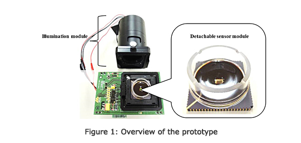
Toshiba has developed a fluorescent imaging system able to observe cells cultured directly on a CMOS image sensor(Note 5) and obtain images that distinguish a single cells in the cluster without an objective lens. Key to this was development of a custom filter able to block illuminated light in defined wavelength, which is deposited directly on the CMOS image sensor (Fig. 1).
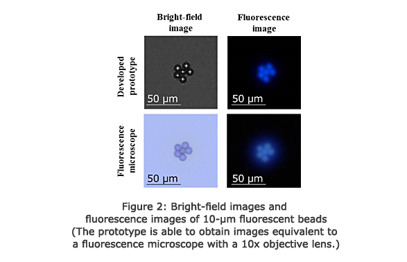
Similar previously proposed technologies have required a computer to process and enhance image resolution in order to distinguish typical cell sizes of around 10μm. Toshiba's system achieves both good filter performance and high spatial resolution, which are usually trade-off against one another, by optimizing filter design. This has improved spatial resolution to the world finest(Note 4) level of less than 10μm, a third of previous values (Fig. 2). The company has also successfully used the fluorescence imaging system inside an incubator to capture fluorescence images of cell clusters(Note 6) in which the nuclei of the target cells were stained.
Since the prototype can capture and send fluorescence images on demand by culturing cells on the sensor module, it has made anytime, remote observation of the state of the cells in the incubator much easier(Note 8) . Furthermore, as the system is small(Note 7), it offers excellent portability and can even be used outdoors.
Future Development, Plans, and Goals
Toshiba is continuing research and development to further improve the value that the fluorescence imaging system will provided to customers, and aims to enter into partnerships with partner companies in order to commercialize the system.
Note that part of this work was funded by ImPACT Program of Council for Science, Technology and Innovation (Cabinet Office, Government of Japan). Some experiments using the prototype were conducted with the cooperation of Takeda Pharmaceutical Company Limited.
- (Note 1)
- A system that uses fluorescent dyes or fluorescent proteins to visualize particular structures or the expression of particular proteins in a cell sample.
- (Note 2)
- System for cultivating cell cultures in controlled conditions of temperature, humidity, and CO2 concentration.
- (Note 3)
- Comparison with methods that perform imaging in a single capture without using special image-processing techniques, such as resolution enhancement. Investigated by the company in April 2017.
- (Note 4)
- Spatial resolution finer than 10μm was confirmed in tests using fluorescent beads.
- (Note 5)
- Known as on-chip imaging or lens-less imaging.
- (Note 6)
- Experiments were conducted on rat primary neurons and human colon cancer cells by using Hoechst® 33342 dye.
- (Note 7)
- The entire system of the prototype is 140 (W) × 70 (D) × 140 (H) mm3 (excluding the PC).
- (Note 8)
- Images of cells placed on the sensor are captured, eliminating the need for an objective lens that must be adjusted and focused.