
Toshiba novel method for quantifying cerebrospinal fluid flow dynamics uses magnetic resonance image analysis
Overview
Toshiba has developed a novel method for automatic computation of cerebrospinal fluid (CSF) flow velocity that uses advanced image processing techniques on non-contrast enhanced magnetic resonance (MR) images. This method can improve the diagnosis of CSF-related conditions, including dementia caused by idiopathic normal-pressure hydrocephalus (iNPH).
Development background
Dementia, a neurodegenerative disease with significant socio-economic impacts, is characterized by impaired memory and decision-making functions. The probability of its onset increases with age, and an estimated 10% of people develop the disease. One known cause of dementia is iNPH, which may be reversible with proper treatment. Time-SLIP (Time-Spatial Labeling Inversion Pulse), a non-contrast enhanced MR imaging technique for visualizing blood flow and CSF flow, can aid in the diagnosis of CSF-related conditions such as iNPH. However, physicians have been limited to qualitatively and manually assessing CSF flow, as technologies for quantitative assessment were unavailable.
Features of the proposed technology
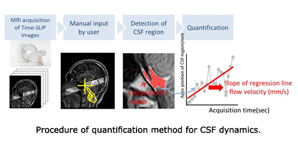
Toshiba was concerned to address this issue, and working in cooperation with Toshiba Medical Systems Corporation and Toshiba Rinkan Hospital has developed an automated method for quantifying CSF flow dynamics, using pixel based time profile analysis of Time-SLIP images. The method has been validated with healthy volunteers, and confirmed a promising 0.91 correlation between actual and measured CSF flow velocity in the prepontine cistern, a diagnostic region for CSF-related diseases. Toshiba’s new automated technology minimizes physician burdens and achieves previously unavailable quantitative assessment of CSF flow dynamics.
Future prospects
Building on the success of the current work, Toshiba will aim to improve quantification accuracy for a wider range of images, including clinical images of normal and diseased regions.