
Corporate Research & Development Center
Development of a technology for automatic localization of the cardiac area and diaphragm from a chest 3-D MRI
Overview
Toshiba, together with Toshiba Medical Systems Corporation, has developed a technology which automatically localizes the cardiac area and diaphragm from a 3-D MRI image of the entire chest area, and confirmed its accuracy in cooperation with Kyorin University Hospital. The technology eliminates complicated procedures required in coronary artery examinations such as determining the location of the heart. This technology was presented at the 16th Annual Society of Cardiovascular Magnetic Resonance (SCMR) Scientific Sessions, the world’s largest conference on cardiac magnetic resonance imaging, held in San Francisco, U.S.A.
Challenges faced by conventional technologies
Cardiac MRI allows doctors to simultaneously examine various heart diseases such as cardiac infarction and cardiomyopathy. During a MRI coronary artery examination, operators must first localize the cardiac area based on an image of the patient's chest in order to re-position the heart near the center of MRI gantry for subsequent magnetic field correction. Since the coronary artery examination requires respiration-synchronized images, the apex of the diaphragm above the liver must also be localized for monitoring respiratory motion. These procedures are conventionally conducted manually, but accurately localizing the cardiac area and diaphragm apex is extremely difficult and imposes a substantial burden on MRI operators.
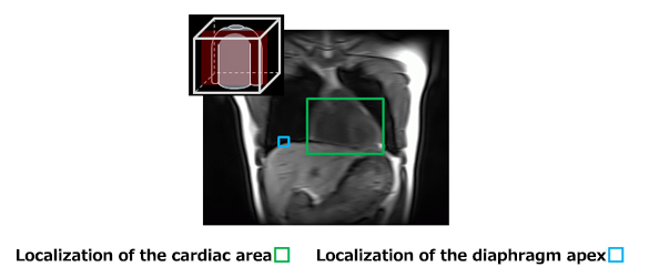
Localization of heart area and diaphragm apex
To solve this issue, Toshiba developed an image alignment technique which aligns a low-resolution 3-D image of the patient's chest area with a model image of a human with an average build. The technology automates the manual localization processes previously required for a coronary artery examination. The model image contains accurate information as to the cardiac area and diaphragm apex location, and can be overlapped with the image of the patient to identify the cardiac area and diaphragm apex. We use a non-rigid image distortion technique to accurately align the patient image with the model image. Generally, non-rigid alignment of 3-D images takes a long time to calculate, but we have developed an algorithm for high-speed image alignment which allows the calculation to be completed in just two seconds on a general-purpose PC. In a joint study with Kyorin University Hospital, we tested this technology on 32 patients with heart diseases and confirmed that the cardiac area and diaphragm apex were localized with sufficient accuracy for diagnosis.
Outlook
Toshiba has established a fundamental technology for automated MRI coronary artery examinations. This automation technology is applicable not only to coronary artery examinations but to all cardiac MRI examinations which require cardiac positioning. The technique makes work easier for operators and reduces examination time so that patients can undergo examinations more comfortably. We aim to further improve alignment accuracy by including clinical data of non-Japanese patients and commercialize a product which utilizes this technology.