
- Back to the previous page
- AI Technology
Myocardial Contour Detection and Cardiac Slice-Alignment Methods for Medical Image Processing
For the accurate evaluation of myocardial perfusion on computed tomography (CT) images, Toshiba has developed a myocardial contour detection method based on an active shape model with highly accurate left ventricle coordinate system estimation. This method eliminates the variations of heart position, rotation, and scale in volume, and thus achieves accurate and fast myocardial contour detection sufficient for myocardial perfusion examination.
This technology has been incorporated into the Aquilion ONE™ whole-body X-ray CT scanner produced by Toshiba Medical Systems Corporation since July 2011.
In order to simplify cardiac scan planning, we have also developed a sophisticated slice-alignment method for cardiac magnetic resonance imaging (MRI) systems. This method employs knowledge-based recognition technologies combined with image processing technologies, making it possible to detect the cardiac planes more quickly and accurately than with the conventional method. It is also more robust with respect to data from a variety of ethnic groups, achieving highly efficient slice alignment.
We presented a paper on this method at the 28th Annual Scientific Meeting of the European Society for Magnetic Resonance in Medicine and Biology (ESMRMB) in October 2011.
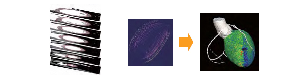
Automatically extracted contour of myocardium of left ventricle (left) and
result of myocardial perfusion (right)
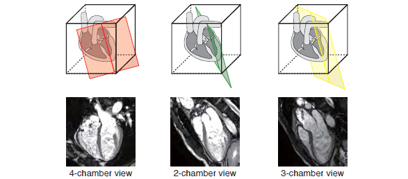
Automatic slice alignment for cardiac MRI